


Knee joint arthrosis is a chronic degenerative (long) disease that causes the destruction of cartilage in the joints.Symptoms include pain, stiffness and swelling.Treatment options to reduce pain and defects include lifestyle changes (diet, physical training), physical and professional treatment methods, medications and surgery.
Osteoarthrosis of the knee joint
Knee joint osteoarthrosis is a common disease, accompanied by chronic and exhausting pain.Recent clinical data shows that central sensitization stimulates osteoarthrosis of the knee joint deformation.A better understanding of how knee joint arthrosis affects the processing of pain centers is important to identify new analgesic targets/new therapeutic strategies.
The barrier receptor weakens the function of the peripheral immune cells and modulates the central neuro-immune response.The systemic introduction of the receptor agonists undermines the pain of OA-caused pain, and circulating and anti-inflammatory changes have been created in this model.
Summarize arthrosis
Arthrosis of the knee joint is inflammation and wears cartilage in the bone that forms the knee joint (osteo = bone, artro = joints, itis = inflammation).Diagnosis of osteoarthritis Knee joint is based on two main results: radiographic data on changes in bone health (using medical images, such as X-ray and MRI images), and human symptoms.About 14 million people have knee arthrosis symptoms.Although more common in older people, 2 million out of 14 million people with knee symptoms are younger than 45 years of diagnosis, and more than half younger than 65 years.
Osteoarthritis (Knee OA) is a progressive disease caused by inflammation and deterioration of the knee joint, which over time increases.
This affects the entire joint, including bones, cartilage, ligaments and muscles.Its development is affected by age, body mass index (BMI), bone structure, genetic, muscle strength and activity level.The OA knee can also develop as a secondary country after a knee injury.Depending on the level of the disease and the presence of the injury or the condition associated with it, the OA knee can be controlled using physical therapy.Worse or developing cases may require surgical intervention.
Symptom
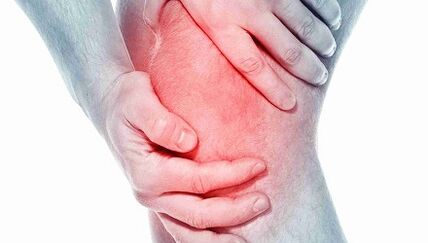
People who develop knee OA may experience a variety of symptoms and restrictions based on the development of the disease.Pain occurs when the cartilage that covers the bone -the knee joint is used.The area where the cartilage is wearing or damaged, exposing the underlying bone.Bone effects allow you to increase the pressure and compression of cartilage, and sometimes bone relationships while moving, which can cause pain.Because the knee is joints, activity levels, activity levels, as well as the type and duration of action, as a rule, have a direct effect on symptoms.Symptoms can decline with heavy activity, for example, when walking with heavy objects.
Symptoms of knee joints may include:
- The deterioration of pain during or after surgery, especially when walking, climbing, walking down the stairs or moving from the seat to a standing position.
- Pain or stiffness after sitting with a knee bent or straight for a long period of time.Pain is the most common symptom of osteoarthritis.When the disease develops and inflammation, the pain can be constant.
- The feeling of jumping out, cracking or grinding when moving the knee.
- Swelling after action.
- The rigidity of the affected joints is often seen for the first time in the morning and after the break.
- Edema, which is sometimes warm to the touch, can be seen in joints with arthritis.
- Deformation can occur with osteoarthritis due to bone growth and cartilage loss.Bone growth in the final joints of the finger is called hyberden node.Bushar nodes are bone growth in the middle of the finger.Deegeneration of cartilage can cause external curvature (onion).
- Crackering or scarring sensation can be observed when arthritis moves.This is caused by sweeping the bones against the bone or the rough cartilage.
Usually these symptoms do not arise suddenly and at once, but gradually develop over time.Sometimes people do not admit that they have osteoarthritis, as they cannot remember the specific time or injury that causes their symptoms.If the knee pain has declined for several months, which does not respond to rest or changes in the activity, it is best to seek advice to medical workers.
Diagnostics
Osteoarthritis can often be diagnosed by symptoms of pain, reduced movement and/or deformation.Osteoarthritis can be confirmed by X-ray or MRI scans.General data include narrowing of the articular space between bones, cartilage loss and spear or bone growth.Blood tests can be used to exclude other possible conditions, but they cannot diagnose osteoarthritis.
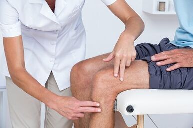
In the knee OA, 2 main processes are diagnosed.The first is based on reports on symptoms and clinical examinations.Physiotherapists will ask about medical history and activities.The therapist will conduct a physical examination to measure the movement of the knee (various movements), strength, mobility and flexibility.They can also ask for various movements to see, improve or reduce pain.
The second tool used to diagnose the knee joint is diagnostic visualization.Physiotherapists can send to a doctor who will prescribe knee X -Rays in various positions to check damage to bone and cartilage.
If more serious damage to the joints is suspected, you can order MRI to be more careful to study the overall status of the articular fabric and its surroundings.
Blood tests can also be ordered to help exclude other conditions that can cause symptoms similar to osteoarthritis of the knee joint.
Treatment
Depending on the severity of arthritis and age of the patient, it will be selected how to treat knee joint arthrosis.Treatment may consist of operating or conservative methods, or their combination.
The first line of treatment of knee joint arthritis includes modification of activity, anti -inflammatory drugs and weight loss.
The rejection of pain that increases pain can make this condition acceptable to some people.Anti -anti -inflammation helps reduce inflammation that can contribute to pain.
Physical therapy to strengthen the muscles around the knee can help absorb some of the shock given to the joints.This is especially true for arthritis with knee cups (patello-femoral).Special types of braces designed to transfer loads to the knee joint, which are less than arthritis, can also relieve pain.Medicines in the knee joint can also help temporarily.
In addition, walking with a stick on the other hand, as painful knees can help distribute some of the burden, reducing pain.Finally, weight loss helps reduce the force through the knee joint.The combination of these conservative steps can help relieve pain and prevent defects.
If this method does not allow you to create a tolerant condition, surgery may be the best option for treating knee joint arthritis.The right type of operation depends on age, anatomy and major conditions.Some examples of surgical options for treating arthritis include osteotomy, which consists of cutting bones to align the joints.
Modern methods of treating knee joint arthrosis include osteotomy, which is a good alternative if young patients and arthritis are limited by a knee joint area.This allows the surgeon to rebuild the knee to unload the arthritis area and carry out the relative load does not involve the knee joint.For example, patients can be rebuilt to redistribute the load through the joints.The advantage of this type of surgery is that the patient's own knee joint is preserved and potentially ensuring years -years of relieving pain without prosthetic knee deficiency.Its weaknesses include longer recovery courses and the possibility of developing arthritis in the new knee.
Operations to replace the knee joints include cutting arthritis bones and prosthetic joint insertion.All surfaces of the arthrit are replaced, including the femur, the lower leg and the knee cup.The arthrit surface is removed, and the ends of the bone are replaced by the prosthesis.The prosthetic components are usually made of metal and plastic surfaces, designed to glide smoothly with each other.
Replace the knee joint
The overall operation to replace the knee joint was first performed in 1968 and for years -years developed in a reliable and effective way to eliminate pain while deadly and allowing patients to continue their active life.Improvements in the field of surgical and implant methods help make this one of the most successful orthopedic procedures today.As the population becomes older and is still more active, the need for general replacement of the knee continues to grow.Many operations to replace the knee joint occur in a special surgical hospital.Improvements in new surgical technology and implant design are some of the donations made by the surgeon.
People often wonder when and why they should replace their knees.This is an individual question that depends on the level of human activity and functional needs.Many people with arthrosis live in pain, which prevents them from participating in activities;The others are so weak that it is difficult for them to wear shoes and socks.Complete replacement of the knee joint offers solutions to arthrosis problems and is done to relieve pain and resume activity.After recovery from the complete replacement of the knee joint, the patient may expect surgery, without pain.Complete replacement of the knee joint improves the patient's condition, and significantly reduces the cost of treatment.This study shows that not only the overall replacement of the knee joint is economically effective, but also provides greater functionality and the best quality of life.
The complete replacement of the knee joint is considered the main operation, and the solution is not trivial.Usually people decide to undergo surgery when they feel that they can no longer live with their arthritis.
Implants consist of 4 parts: tibia, femoral parts, plastic inserts and patterns.The tibia and femur components are made of metal, usually chromium cobalt, used to close the thighs and lower legs after removing arthritis bones.Plastic inserts are made of polyethylene mass of ultra -high molecules and fit the tibia component, so that the thigh surface is polished along the plastic.The knee cup component also slides forward the femoral component.They are usually attached to bone cement.
Full knee replacement is performed in the operating room with a special laminar airflow system, which helps reduce the likelihood of infection.Your surgeon will wear "space", also designed to reduce the likelihood of infection.The entire surgical team will consist of your surgeon, from two to three assistants and caregivers.
Anesthesia is given through the epidural catheter, which is a small tube that is inserted backwards.During surgery, the patient can be awake and drowsiness.
After the introduction of the epidural block around your thighs, the tourniquet or cuff will be placed.Horizontal bars will be exaggerated during surgery to reduce blood loss.The cut for a complete replacement of the knee is made along the front knee.The incision will be measured from 4 to 10 inches depending on the anatomy.
The surface of the femur arthrit, the lower leg and the patella are exposed and removed using strength tools.At the same time, the knee deformation is corrected, and after surgery, the knee becomes more straight.Bones are ready to take artificial knee joints, and then prosthesis are inserted.During the closure, two drainage is installed around the work area to assist in blood transfusions.Sapers are used to cover the skin.
The entire operation will take 1 to 2 hours.After that, the patient will be taken to the recovery room where the test will be examined.Most patients can be taken to the normal room for several hours;Other people need to stay in the hall for recovery, as defined by surgeons and anesthetists.
The patient usually lives in the hospital for 3-4 days after the full operation to replace the knee
Risk during surgery
Some risks of surgical procedures include blood loss, leg formation and the probability of infection.The generalization of this risk is very small.They should be discussed with the surgeon before the start of the operation.
Some risks of prosthetic knee presence include the possibility that the parts may weaken or worn over time, or the prosthesis may be infected.Again, these issues will be discussed with the surgeon.
Post -operative course
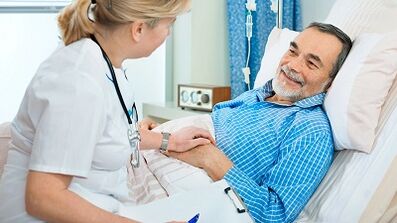
Immediately after a complete operation to replace the knee joint, the patient will fall into the recovery room.Most patients can enter the normal ward after a few hours, when the sensation returns to the feet.The pain pump associated with the epidural catheter will be given, which will allow you to control when the antidote for pain is given.
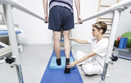
On the day of surgery, you can do some exercises, as shown by physiotherapists, including reduction in quadriceps and moving their feet up and down.Depending on the priority of the surgeon, you can start bending your new knee as soon as the operation or on the first day.Patients will be allowed to take ice after surgery to wet their mouths, but drink fluids or you can cause nausea.Patients will have a catheter in the bladder, so you don't have to worry about urination.Once the foot movement is restored, it will be allowed to sit, get up and take a few steps with a walker and therapy.
The first day after operation will be active, developed to help to be more mobile.
The patient will meet with a physiotherapist who will direct additional training.In addition, they will help to get their feet and take a few steps on foot.As a rule, the patient will be allowed to drink pure fluid.
In the next few days, it's easier and easier to move.The patient will be released from pain and urinary catheter.Pain treatment will be given in the form of tablets.On the second day after surgery, if the signs of recovery are found in the intestine, it will be allowed to eat regular foods.
Depending on your age, pre -operation and insurance coating, patients may be candidates for short accommodation in rehabilitation institutions.Otherwise, the patient will be released home, and the physiotherapist will come to his home to continue his recovery.The sender will discuss this option with the patient and will help him plan his home.
Back to the activity will be guided by the surgeon and therapist.As a rule, patients can walk as much as they want 6 weeks after surgery.Patients can continue movement after 6 weeks.After 8 weeks, the patient can continue the game in golf and swim;At 12 weeks they can play tennis.The surgeon will help determine the action that can be resumed.
What does the physiotherapy need
All physical therapists are provided through education and clinical experience for the treatment of various conditions or injuries:
- A physiotherapist with experience treating people with osteoarthritis of the knee joint and after surgery to replace the knee joint.Some physiotherapists have the practice of orthopedic focus.
- Physiotherapist who is a certified orthopedic clinical specialist.These physiotherapists will have sophisticated knowledge, experience and skills that can be used for the situation.
- You can find physiotherapists who have these accounting data and others using MRI, online tools to help find physiotherapists with certain clinical knowledge.
General advice when you can find a physiotherapist (or any other medical service provider):
- Get suggestions from family and friends or from other medical services providers;
- Switching to the clinic for physiotherapy for admission, you need to ask about the experience of physiotherapists in helping people with arthritis.
During your first visit with a physiotherapist, you need to be prepared to describe the symptoms as more detailed, and report on activities that worsen the situation.